Atrial fibrillation is the most common treated heart arrhythmia. A-fib is a nickname for this common condition.
To identify atrial fibrillation on an ECG strip, you first need to understand how the P wave and QRS complex are developed.

A normal heart atrial contraction will show a normal P wave on the ECG.
Contraction of ventricles is associated with QRS on the ECG.
Usually, each heartbeat starts in the atrium and then travels into the heart's ventricles, which translates on the ECG into a P wave before each QRS.
Now that you understand the normal mechanism of the heart's electricity, let's see what can go wrong.
In the case of atrial fibrillation, the heart starts generating chaotic noncoordinated impulses from multiple abnormal regions within the atrium, which we call ectopic electrical discharges.
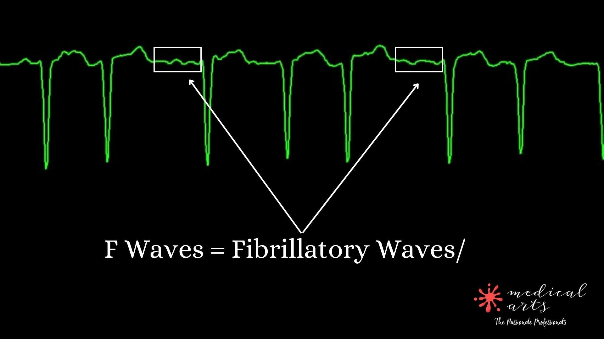
This abnormal atrial behavior translates on the ECG as non-existent P waves, but only F waves, which refers to fibrillatory waves. This condition makes the atrial contractions insufficient to trigger heart contractions.
This results in irregular heartbeats that can be seen on the ECG as the irregular distance between R-R waves.
To recap in one sentence, Atrial fibrillation is distinguished by irregular heartbeats and absent P waves.
Slow Atrial fibrillation is harmless, but if the atrial fibrillation has a heart rate faster than 100 beats per minute, it needs to be treated to avoid complications such as strokes.