The human foot is a remarkable structure comprising numerous bones, joints, and muscles that enable us to stand, walk, run, and perform various activities.
Overview of the Foot Bones
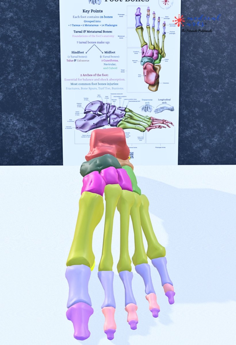
The foot is divided into three main regions:
the tarsal bones,
the metatarsal bones,
and the phalanges.
Each of these regions plays a crucial role in the foot's function and mobility.
1. Tarsal Bones
The tarsal bones form the posterior part of the foot and are responsible for the foot's stability and movement. There are seven tarsal bones:
- Calcaneus: This is the largest bone in the foot, forming the heel. It is the primary weight-bearing bone and supports us when we stand or walk.
- Talus: Positioned above the calcaneus, the talus connects the foot to the lower leg. It plays a critical role in the ankle joint, allowing for the up-and-down motion of the foot.
- Navicular: Located in front of the talus, the navicular bone helps connect the tarsal bones to the metatarsal bones.
- Cuboid: Found on the foot's outer side, the cuboid bone is essential for lateral stability and supports the foot's arch.
- Cuneiform Bones: There are three cuneiform bones:
- Medial Cuneiform: Located on the inner side of the foot.
- Intermediate Cuneiform: Situated between the medial and lateral cuneiform bones.
- Lateral Cuneiform: Found towards the outer side of the foot.
2. Metatarsal Bones
The metatarsal bones are the long midfoot bones connecting the tarsal bones to the phalanges. There are five metatarsal bones, each corresponding to a toe:
- First Metatarsal: Supports the big toe (hallux).
- Second Metatarsal: Supports the second toe.
- Third Metatarsal: Supports the third toe.
- Fourth Metatarsal: Supports the fourth toe.
- Fifth Metatarsal: Supports the little toe.
These bones are crucial for weight distribution and balance during movement.
3. Phalanges (Toe Bones)
The phalanges are the bones that make up the toes. Each toe has three phalanges, except for the big toe, which has only two. The phalanges are divided into three types:
- Proximal Phalanges: The bones closest to the metatarsals.
- Intermediate Phalanges: The middle bones (absent in the big toe).
- Distal Phalanges: The bones at the tip of the toes.

Joints of the Foot
The foot's bones are connected by numerous joints that allow for a wide range of motion:
- Ankle Joint: Formed by the talus, tibia, and fibula, this joint allows the foot to move up and down.
- Subtalar Joint: This joint enables side-to-side motion between the talus and calcaneus.
- Transverse Tarsal Joint: Connects the tarsal bones and allows twisting movements.
- Tarsometatarsal Joints: Connect the tarsal bones to the metatarsal bones.
- Metatarsophalangeal Joints: Connect the metatarsals to the proximal phalanges.
- Interphalangeal Joints: Connect the phalanges (proximal to intermediate and intermediate to distal).

Functions of the Foot Bones
The foot bones perform several critical functions:
- Support: The foot bones support the body's weight when standing, walking, or running.
- Movement: The arrangement of bones and joints allows for complex movements, including walking, running, jumping, and balancing.
- Shock Absorption: The arches of the foot, supported by the tarsal and metatarsal bones, help absorb impact and reduce stress on the body.
- Stability: The foot's structure provides a stable base for the body, helping to maintain balance and posture.
Common Foot Bone Injuries
Understanding foot anatomy is essential for recognizing common injuries, which include:
- Fractures: Breaks in any foot bone, often caused by trauma or overuse.
- Sprains: Stretching or tearing of ligaments, typically affecting the ankle joint.
- Plantar Fasciitis: Inflammation of the tissue connecting the heel bone to the toes, causing heel pain.
- Bunions: A bony bump that forms on the joint at the base of the big toe.
Conclusion
The anatomy of foot bones is a fascinating and complex subject, integral to our daily activities and overall mobility. By understanding the structure and function of these bones, students and the general public can better appreciate the importance of foot health and recognize potential issues that may arise. Whether for educational purposes or general knowledge, this overview provides a solid foundation for further exploration into the remarkable world of foot anatomy.